A Neurobiological Consideration of Humor
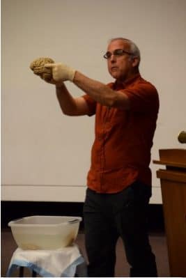
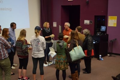
Prof. White recently participated in a unique interdisciplinary conference on humor at the University of North Carolina Chapel Hill. His talk and demonstration was entitled, “A Neurobiological Consideration of Humor”. In keeping with his Medical Neuroscience style, he brought human brain specimens to demonstrate to the diverse audience how the brain processes humor. The audience included, social scientists, psychologists, philosophers, humanists, feminists, humorists, television “sit-com” writers and producers, professional comedians, and students from a variety of disciplines. Several attendees gathered around the brains at the conclusion of the presentation for a closer look, to ask questions, and to hold a brain.
Neurobiology of Humor
The journalist of “Learn Medical Neuroscience” lives in the Netherlands and could not be present at Prof. White’s presentation. For the information on Neurobiology of Humor the publication: “Humor Modulates the Mesolimbic Reward Centers” (2003), Dean Mobbs et al., Neuron, Vol. 40 Issue 5. has been used.
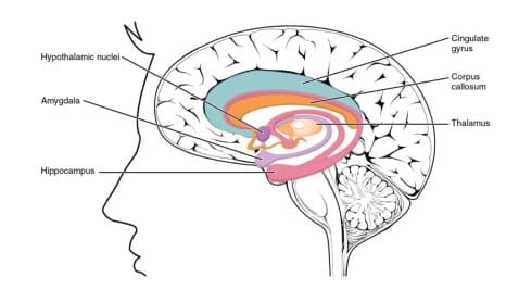
Humor plays a very important role in many facets of human life including psychological, social, and somatic functioning. Humor modulates activity in several cortical regions, and there is evidence that it engages a network of subcortical regions including the nucleus accumbens (part of the basal ganglia, the ventral tegmental area (located in the midbrain) and amygdala, key components of the mesolimbic dopaminergic reward system. The authors found that funny cartoons when contrasted with nonfunny cartoons activated the temporo-occipital junction, IFG (Inferior Frontal Gyrus)/temporal pole, and SMA (Supplemenrary Motor Area) /dACC (dorsal Anterior Cingulate Gyrus), all in the left hemisphere.
In their study the largest area of cortical activation occurred in the left lateral IFG, including Broca’s area, possibly reflecting the language-based decoding of the stimuli. In the literature IFG is described is as a polymodal language region, involved in numerous aspects of language processing, including semantic and sentence processing. The role of the left SMA proper and pre-SMA are likely to reflect motor aspects of expressive laughter. The amygdala, ventral striatum/nucleus accumbens, ventral tegmental area, anterior thalamus, and the hypothalamus form the core of the subcortical dopaminergic reward network.