by: Leonard E. White, PhD, Durham NC (U.S.A.)
The next session of Medical Neuroscience begins today (August 29, 2016). We look forward to welcoming to our course another group of wonderfully diverse and passionate learners from all walks of life and all over the world. One particular corner of the world that will contribute some number of students to the course is Durham North Carolina (U.S.A.), home to Duke University.
Neuroscience Bootcamp
Today, future neuroscientists who matriculated at Duke University this summer begin their doctoral studies in an intensive two-week course we call Neuroscience Bootcamp. This “bootcamp” begins with an afternoon in the teaching laboratory of the Duke Institute for Brain Sciences. There I have the privilege of introducing these students to the anatomy and function of the human brain. There are 22 students in this group who begin their 5-7 year journey toward completion of a PhD in one of several graduate programs we offer here at Duke. Their dissertations will explore the diverse interests of our faculty which span the field from molecular mechanisms of synaptic plasticity and computational neuroscience to brain tumor biology and cognitive neuroscience. It truly is a wonderful opportunity these students have to prepare for careers in the brain sciences or in some vocation that applies neuroscience discovery to improve the human condition.
Functional Anatomy of the Human Brain
These new graduate students are not the only Duke students to join us online today. I also begin a fall semester course that I teach for baccalaureate students at Duke University called “Functional Anatomy of the Human Brain”. This course will have 24 students, most of whom are senior undergraduate students majoring in neuroscience. This course was originally designed for 16 students, but over the last few years I increased the capacity to accommodate strong student interest. Despite growing the course to 24, I still have 18 students on a “wait list” that would be eager to join the course. I wish I could accommodate all interested students on campus the way we can online!
Blended learning at Duke University
For both of these groups of students, I will encourage enrollment in Medical Neuroscience as a way for students to prepare for what we do in the classroom and laboratory.
Thus the students wil be in a blended learning course. Blended learning is a formal education program in which a student learns at least in part through delivery of content and instruction via digital and online media with some element of student control over time, place, path, or pace. (information on blended learning added by Ellen Vos).
This will be a consistent experience for my baccalaureate students throughout the fall semester. They will work through lessons in Medical Neuroscience assigned to support the learning I have planned in the campus-based course. My goal is to give these students multiple means for preparing themselves for what we come together in class to accomplish. Today, the goal is to find the central sulcus in the human brain and to learn about how the body is represented along the length of the precentral and postcentral gyri. I am hoping that my students will accomplish with brains in their hands what I demonstrated in the Week 2 video, “Finding the Central Sulcus”. That seems to me like a great way to begin a course on functional neuroanatomy!
Research on somatic sensory and motor structures in the human nervous system
This topic takes me back to some research I conducted with Dale Purves back in the mid-1990s. We studied the morphology of somatic sensory and motor structures in the human nervous system. With particular interest in the possibility that cerebral asymmetry might explain handedness. During the course of this study, we were quite surprised to discover just how variable the central sulcus can be among brains, and even within the two hemispheres of the same brain. Evidently, the editors of the journal (Cerebral Cortex) were impressed as well, since they chose to put our Figure 2 on the cover of the journal issue that presented our two, back-to-back papers.
- White LE, Andrews TJ, Hulette C, Richards A, Groelle M, Paydarfar J, Purves D (1997a). Structure of the human sensorimotor system I. Morphology and cytoarchitecture of the central sulcus. Cerebral Cortex 7:18-30.
- White LE, Andrews TJ, Hulette C, Richards A, Groelle M, Paydarfar J, Purves D (1997b) Structure of the Human Sensorimotor System II. Lateral symmetry. Cerebral Cortex 7:31-47.
Variations in the brain
Below is an image showing that figure from White et al. (1997), Cerebral Cortex 7:18-30. I cut out of the same post mortem human brains (8 shown below) blocks of cerebral hemisphere containing the central sulcus in the two hemispheres. After photographing these blocks and scanning their photographs (before the days of digital photography), I colorized the precentral gyrus red and the postcentral gyrus blue. Even today, I remain impressed at how variable this part of the human brain can be from one person to the next. Vive la difference!
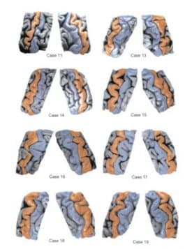
Although we failed to find compelling evidence favoring a cerebral asymmetry that might explain handedness in this study, other investigators in the years following used MRI methods and much larger samples to suggest that indeed, “bigger might be better” when it comes to the representation of the preferred hand in the human brain.
Welcome
Hope you enjoy your continued journey of learning and discovery in Medical Neuroscience. Now with a new cohort of eager students joining from Duke University and from universities, libraries, cafés, and homes all around the world!